Flow changes
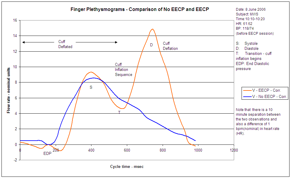
The ideas discussed in previous posts are quite difficult to envision but it helps to check the diagrams produced by the plethysmograph. Remember that the instrument measures the blood flow through a finger during the cycle of a heartbeat. Recall that the therapist uses these traces to adjust the EECP unit for best results with each individual. The data on the chart are taken from one of the author’s sessions; they have been re-traced to a common timebase and there are minor errors in the graphs due to time and zero differences. The flow differences are, however, quite striking through the cycle.The blue trace shows the flow pattern when the body is at rest and the orange trace while undergoing EECP.
Heartbeat cycle
Detailed accounts of the heart’s operation can be found in many sources so what follows only covers those aspects to help understand EECP. If you are a heart patient you will doubtless have heard the terms systolic and diastolic blood pressures. These are the maximum and minimum pressures in the measured artery during the heartbeat cycle. The words are derived from ‘systole’ and ‘diastole’ – of Greek origin meaning ‘contraction’ and ‘expansion’ respectively. They refer to the pumping and relaxation phases of the cycle.
Referring to the chart, we start with the heart being fully relaxed, the lower chambers (atria) being full of blood (marked EDP – End Diastolic Pressure). In the systolic phase the heart muscle is compressing, increasing the pressure and squeezing the blood out of the left and right upper chambers of the heart, the ventricles. The left ventricle blood goes through the open aortic valve to the aorta, generating the maximum (systolic) pressure (marked S – Systole) As the left ventricle empties so the contraction phase finishes, the pressure drops around the valve and it shuts. On the blue curve the pressure then reduces fairly evenly until the next systolic phase begins.
Enhanced flow
With EECP, however, the system is looking for the closure of the aortic valve by interpreting the ECG waveform produced by the heart. It does not have to do this exactly because the therapist can control in detail the start point of leg cuff inflation to obtain the best results. The start of inflation is called the Transition (T) and, on the orange trace, the increase in flow after that point is obvious.
The process of inflation itself is a bit more subtle; the reason that there are three sets of cuffs is that the inflation time for each cuff can be sequenced, the lowest cuff being inflated first, followed by the two upper ones in sequence, each about 50 msec (1/20 second) after the other. In effect the blood is chased back to the heart rather like an extremely rapid squeeze along a toothpaste tube. This process drains the leg of blood and so, as the air pressure is then released simultaneously, the leg tissues expand elastically allowing the blood to flow back with less pumping effort from the heart muscle. The result is that the end diastolic pressure (EDP) is typically lower than in normal operation. The same elastic rebound effect is true at systole, leading to an overall lower load on the heart muscle during EECP.
The therapist and the patient are both interested in the shape of the enhanced diastolic flow curve, the patient in the hope of getting the best from the EECP procedure and the therapist in achieving that goal. The enhancement is typically measured by both the peak value (P) – the maximum flow in comparison with normal operation – and the total area under the curve (A). The area represents the total increase in flow of blood caused by EECP. The greater this is, the more oxygenated arterial blood flows through the coronary arteries.
EECP Effects on the body
One of the most interesting aspects of EECP is that the blood flow effects occur not only in the finger with the plesymograph sensor and in the coronary arteries, but in practically every blood vessel in the body. Thus any disease or condition of the body ‘/affected by impaired blood flow may potentially be improved by a course of EECP. In the author’s case a nagging arthritic joint of the right big toe that had been troublesome for at least 5 years has miraculously improved so that there is now no pain. The present state of knowledge is such that the medical profession cannot make a firm promise of such benefits and thus one should regard them as a real bonus, should they occur.
Effects on the Heart
Heart disease is not simply a question of blockages in certain identifiable locations. If you have a blockage in one or more of your coronary arteries, what do you suppose is the state of the capillaries joined to those arteries? In other words one must expect partial or complete blockage of the blood supply system. Bluntly, the difference between the living and the dead is that the living still have enough open pathways for the heart to function. Heart disease is therefore a system problem. The hopeful part of this for EECP patients is that the healing factors should work on all parts of the blood distribution system, not just the identifiable coronary arteries. Atheroma in the capillaries (the microvascular circulation) can equally cause angina because of the shortage of oxygen supplied to that area of the muscle. The prospective EECP patient should, almost literally, take heart from the prospect that the treatment will improve the whole system, not just the areas of damage identified by your consultant.
Surface and flow effects
A good part of this section is informed speculation as to what happens in the coronary artery circulation and the possible effects that occur. Also this is written by an engineer, rather than a medic, so some of the understanding may be suspect and open to review by wiser and more knowledgeable types!
As already noted in this thread, engineers get very interested when pressure waves occur in tubes because of the sometimes unexpected effects. However, they are normally used to tubes that are the same diameter along their length and do not move about very much. A blood vessel is a very different proposition. Not only does it change in diameter while flow is occurring but it can move substantially in space, bending and flexing as the diameter is changing. To add more complication, blood itself is not a normal fluid, its viscosity – the readiness to flow - lessens the faster it flows. It is called a non-Newtonian fluid, named after you-know-who. So, to analyse in detail how the flow varies during a heartbeat cycle becomes a very complicated exercise. One way of understanding the interplay of these factors is to develop a model, usually as a computer program, where the physical laws governing the flow and the geometric shape of the artery are used to calculate the effects of various disturbances including progressive atheromas. While this work has been under way for a few years it does not yet appear to be advanced enough to provide real guidance to clinicians.
To add to the complication blood vessels possess a very special surface and wall. The surface or inner lining is called the endothelium. Backing up the endothelium and giving strength to the wall is a layer of smooth muscle. The endothelium possesses almost mystical properties that have a fundamental effect on the operation of the blood vessel, in particular the expression and absorption of certain chemical substances that regulate the operation of the smooth muscle. If the endothelium does not work properly then the blood vessel can be badly affected. Atheroma tends to block the vessel by accumulating a hard fatty deposit (plaque) under the surface, expanding into the vessel and, in the worst case , erupting into the vessel and causing a complete blockage further downstream.
The chemical substances of interest to this discussion (there are many others) fall into several categories:-
- Growth Factors
- Vaso-dilators
- Vaso-constrictors
- Inflammatory agents
- Anti inflammatory agents
Vaso-dilators and vaso-constrictors control the diameter of the blood vessel by acting on the smooth muscle underneath. Growth factors control the production of new micro-arteries (angiogenesis) and the bringing back into service of existing arteries (recruitment)that have ceased to function for a variety of reasons including atheroma.
If the balance between these substances goes wrong for any reason then the consequences can be far-reaching. Endothelial dysfunction[i] is the general name for these problems.
[i] Endothelial Functions: Cardiac Events. Lerman & Zeiher. Circulation 2005;111;363-368
